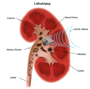
Lithotripsy is a noninvasive (the skin is not pierced) procedure used to treat kidney stones that are too large to pass through the urinary tract. Lithotripsy treats kidney stones by sending focused ultrasonic energy or shock waves directly to the stone first located with fluoroscopy (a type of X-ray “movie”) or ultrasound (high frequency sound waves). The shock waves break a large stone into smaller stones that will pass through the urinary system. Lithotripsy allows persons with certain types of stones in the urinary system to avoid an invasive surgical procedure for stone removal. In order to aim the waves, your doctor must be able to see the stones under X-ray or ultrasound.
The introduction of lithotripsy in the early 1980s revolutionized the treatment of patients with kidney stone disease. Patients who once required major surgery to remove their stones could be treated with lithotripsy, and not even require an incision. As such, lithotripsy is the only non-invasive treatment for kidney stones, meaning no incision or internal telescopic device is required.
Lithotripsy involves the administration of a series of shock waves to the targeted stone. The shock waves, which are generated by a machine called a lithotripter, are focused by x-ray onto the kidney stone. The shock waves travel into the body, through skin and tissue, reaching the stone where they break it into small fragments. For several weeks following treatment, those small fragments are passed out of the body in the urine.
In the two-plus decades since lithotripsy was first performed in the United States, we have learned a great deal about how different patients respond to this technology. It turns out that we can identify some patients who will be unlikely to experience a successful outcome following lithotripsy, whereas we may predict that other patients will be more likely to clear their stones. Although many of these parameters are beyond anyone's control, such as the stone size and location in the kidney, there are other maneuvers that can be done during lithotripsy treatment that may positively influence the outcome of the procedure. At the Brady Urological Institute, our surgeons have researched techniques to make lithotripsy safer and more effective, and we incorporate our own findings as well as those of other leading groups to provide a truly state of the art treatment.
Other procedures that may be used to treat kidney stones include:

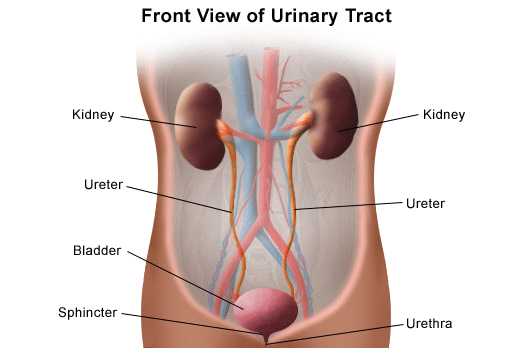
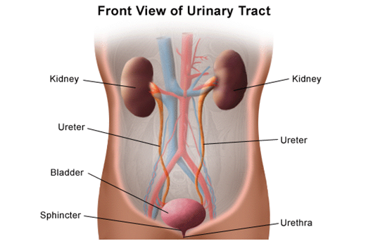
When substances that are normally excreted through the kidneys remain in the urinary tract, they may crystallize and harden into a kidney stone. If the stones break free of the kidney, they can travel through, and get lodged in, the narrower passages of the urinary tract. Some kidney stones are small or smooth enough to pass easily through the urinary tract without discomfort. Other stones may have rough edges or grow as large as a pea causing extreme pain as they travel through or become lodged in the urinary tract. The areas most prone to trapping kidney stones are the bladder, ureters, and urethra.
Most kidney stones that develop are small enough to pass without intervention. However, in about 20 percent of cases, the stone is greater than 2 centimeters (about one inch) and may require treatment. Most kidney stones are composed of calcium; however, there are other types of kidney stones that can develop. Types of kidney stones include:

The body takes nutrients from food and converts them to energy. After the body has taken the food that it needs, waste products are left behind in the bowel and in the blood.
The urinary system keeps chemicals, such as potassium and sodium, and water in balance, and removes a type of waste, called urea, from the blood. Urea is produced when foods containing protein, such as meat, poultry, and certain vegetables, are broken down in the body. Urea is carried in the bloodstream to the kidneys.
The kidneys remove urea from the blood through tiny filtering units called nephrons. Each nephron consists of a ball formed of small blood capillaries, called a glomerulus, and a small tube called a renal tubule. Urea, together with water and other waste substances, forms the urine as it passes through the nephrons and down the renal tubules of the kidney.
The primary advantage of lithotripsy is that it is completely non-invasive.
Lithotripsy is well suited to patients with small kidney stones that can be easily seen by x-ray.
When kidney stones become too large to pass through the urinary tract, they may cause severe pain and may also block the flow of urine. An infection may develop. Lithotripsy may be performed to treat certain types of kidney stones in certain locations within the urinary tract.
There may be other reasons for your doctor to recommend lithotripsy.
You may want to ask your doctor about the amount of radiation used during the procedure and the risks related to your particular situation. It is a good idea to keep a record of your past history of radiation exposure, such as previous scans and other types of X-rays, so that you can inform your doctor. Risks associated with radiation exposure may be related to the cumulative number of X-ray examinations and/or treatments over a long period of time.
Complications of lithotripsy may include, but are not limited to, the following:
Contraindications for lithotripsy include, but are not limited to, the following:
Patients with cardiac pacemakers should notify their doctor. Lithotripsy may be performed on patients with pacemakers with the approval of a cardiologist and using certain precautions. Rate-responsive pacemakers that are implanted in the abdomen may be damaged during lithotripsy.
There may be other risks depending on your specific medical condition. Be sure to discuss any concerns with your doctor prior to the procedure.
Obesity and intestinal gas may interfere with a lithotripsy procedure.

Because lithotripsy is a completely non-invasive therapy, most lithotripsy treatments are performed on an outpatient basis.
Although the use of anesthesia does depend on patient and physician preference, recent data suggest that the results of lithotripsy may be improved with the administration of a mild anesthetic.
When the patient has been adequately anesthetized, a computerized x-ray machine is used to pinpoint the location of the stone within the kidney. A series of shock waves (several hundred to two thousand) is administered to the stone. Our treatment protocols incorporate the latest research findings which suggest that adjustments of both the shock wave power and the rate at which the shock waves are delivered can affect treatment outcome.
Our goal when performing lithotripsy is to maximize the breakage of a patient's kidney stone while minimizing injury that the shock waves can cause to the kidney and surrounding organs.
Typically, an lithotripsy procedure lasts for approximately one hour.
Generally, lithotripsy follows this process:
After the surgery you will be taken to the recovery room for observation. Once your blood pressure, pulse, and breathing are stable and you are alert, you will be taken to your hospital room or discharged home.
You may resume your usual diet and activities unless your doctor advises you differently.
You will be encouraged to drink extra fluids to dilute the urine and reduce the discomfort of passing stone fragments.
You may notice blood in your urine for a few days or longer after the procedure. This is normal.
You may notice bruising on the back or abdomen.
Take a pain reliever for soreness as recommended by your doctor. Aspirin or certain other pain medications may increase the chance of bleeding. Be sure to take only recommended medications.
You may be given antibiotics after the procedure. Be sure to take the medication exactly as prescribed.
You may be asked to strain your urine so that remaining stones or stone fragments can be sent to the lab for examination.
A follow-up appointment will be scheduled within a few weeks after the procedure. If a stent was placed during the procedure, it may be removed at this time.
Notify your doctor to report any of the following:
Your doctor may give you additional or alternate instructions after the procedure, depending on your particular situation.